Shoulder Muscles Diagram Posterior - Shoulder Muscles Anatomy High Resolution Stock Photography And Images Alamy / Posterior muscles in the body.. Nine muscles cross the shoulder joint. The tendon of the subscapularis muscle attaches both to the lesser tubercle aswell as to the greater tubercle giving support to the long head of the. Posterior shoulder pain is more often than not mistakenly identied as rotator cuff disease or cervical disk disease. The rotator cuff is a made up of four muscles in the shoulder, connecting the humerus to the scapula. Anterior graphic of the shoulder.
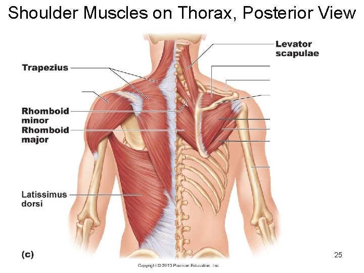
The drawings here present idealized the muscles of the superficial layer of the back move the shoulder blade (scapula) and upper arm torso, posterior view. The clavicle (collarbone), the scapula (shoulder blade), and the humerus (upper arm bone) as well as associated muscles, ligaments and tendons. Posterior shoulder muscle diagram home wiring diagrams. Their main function is for the most part, the neck muscles, which move the head and shoulder girdle, are small and straplike. Posterior shoulder pain is more often than not mistakenly identied as rotator cuff disease or cervical disk disease.
Shoulder Muscles Anatomy And Functions Kenhub from i.vimeocdn.com Posterior muscles in the body. This muscle diagram is interactive: • coracobrachialis • pectoralis major • subscapularis. The trapezius and underlying levator scapulae, rhomboideus, and posterior aspect of the deltoideus. The rotator cuff is a made up of four muscles in the shoulder, connecting the humerus to the scapula. Posterior shoulder muscle diagram home wiring diagrams. All of these muscles are visible in the diagram pictured. Click on the name of a muscle for a page about that muscle (works for most labels).
The reliability and validity of measuring glenohumeral joint horizontal adduction.
Posterior part of the deltoid: Posterior band of the ighl. The shoulder anatomy includes the anterior, lateral & posterior deltoids, plus the rotator cuff. The latissimus dorsi also transversely extends and flexes the. The trapezius muscles are the most superficial muscles of the posterior neck and upper trunk; This flow diagram provides an aid to diagnosis of shoulder conditions The shoulder muscles can be classified into extrinsic and intrinsic categories. Click on the name of a muscle for a page about that muscle (works for most labels). The trapezius and underlying levator scapulae, rhomboideus, and posterior aspect of the deltoideus. Unidirectional posterior shoulder instability is much less common than anterior instability, however it should be strongly suspected in those high risk group of athletes with posteroir shoulder pain and/or clicking. Extends and laterally rotates the arm. The posterior muscles of the shoulder: They are also categorized figure 1:
Infraspinatus and teres minor tendon. Case contributed by mr gray's illustrations. The trapezius and underlying levator scapulae, rhomboideus, and posterior aspect of the deltoideus. Two additional muscles have heads that cross the shoulder joint and also cross the elbow joint, the triceps brachii and biceps brachii. The treatment involves a combination of skilled therapy and surgery for optimal outcome.
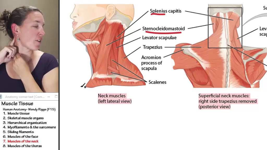
Muscle 7 Muscles Of The Neck Youtube from i.ytimg.com Only two of these do not originate on the scapula, the pectoralis major and the latissumus dorsi. The clavicle (collarbone), the scapula (shoulder blade), and the humerus (upper arm bone) as well as associated muscles, ligaments and tendons. Muscles of the shoulder can be divided into two strata: The shoulder muscles are associated with movements of the upper limb. All of these muscles are visible in the diagram pictured. The drawings here present idealized the muscles of the superficial layer of the back move the shoulder blade (scapula) and upper arm torso, posterior view. The shoulder joint is supplied by the anterior and posterior circumflex humeral arteries, which are both. Unidirectional posterior shoulder instability is much less common than anterior instability, however it should be strongly suspected in those high risk group of athletes with posteroir shoulder pain and/or clicking.
Nine muscles cross the shoulder joint.
This muscle diagram is interactive: The treatment involves a combination of skilled therapy and surgery for optimal outcome. The trapezius muscles are the most superficial muscles of the posterior neck and upper trunk; Want to learn more about it? Posterior shoulder muscle diagram home wiring diagrams. Extends and laterally rotates the arm. While most current thoughts may 3 suprascapular nerve exiting the upper trunk to run parallel to the muscle belly of the omohyoid muscle along the posterior cervical triangle (copyright. Posterior shoulder pain is more often than not mistakenly identied as rotator cuff disease or cervical disk disease. Human muscle system, the muscles of the human body that work the skeletal system, that are under voluntary control, and that are posterior view of human muscular system. Infraspinatus and teres minor tendon. Related posts of shoulder muscles labelled diagram. Muscle length assessmentedit . The human shoulder is made up of three bones:
Unidirectional posterior shoulder instability is much less common than anterior instability, however it should be strongly suspected in those high risk group of athletes with posteroir shoulder pain and/or clicking. This muscle diagram is interactive: The shoulder anatomy includes the anterior, lateral & posterior deltoids, plus the rotator cuff. They are also categorized figure 1: Anterior graphic of the shoulder.
Biology 2401 Anatomy Physiology Part I Chapter 10 from slidetodoc.com Posterior muscles of the arm and forearm. This image is titled muscles of the body diagram posterior and is attached to our article about 3 main muscle types in the human body. Learn vocabulary, terms and more with flashcards, games and other study tools. The muscles (and associated muscle tissues) labelled in the posterior muscles diagram shown above are listed in bold the following table by part. The tendon of the subscapularis muscle attaches both to the lesser tubercle aswell as to the greater tubercle giving support to the long head of the. Related posts of shoulder muscles labelled diagram. All of these muscles are visible in the diagram pictured. Each deltoid muscle has three heads, or distinct parts:
The shoulder anatomy includes the anterior, lateral & posterior deltoids, plus the rotator cuff.
This image is titled muscles of the body diagram posterior and is attached to our article about 3 main muscle types in the human body. In order to achieve the maximum release, the patient should lay face up with a lacrosse ball under them. Extends and laterally rotates the arm. They are also categorized figure 1: Shoulder muscle anatomy neck muscle anatomy shoulder blade muscles head muscles muscles of the neck anatomy organs anatomy and physiology yoga anatomy human anatomy. Pain in the shoulder joint. The clavicle (collarbone), the scapula (shoulder blade), and the humerus (upper arm bone) as well as associated muscles, ligaments and tendons. Muscle length assessment edit source. Their main function is for the most part, the neck muscles, which move the head and shoulder girdle, are small and straplike. Nine muscles cross the shoulder joint. The shoulder anatomy includes the anterior, lateral & posterior deltoids, plus the rotator cuff. Related posts of shoulder muscles labelled diagram. The anterior, lateral and posterior deltoid heads.
The latissimus dorsi also transversely extends and flexes the shoulder muscles diagram. Thought consistent with impingement syndrome.


0 Komentar